
Figure 1. Primary human peripheral blood APCs exhibit spontaneous uptake of plasmid DNA.
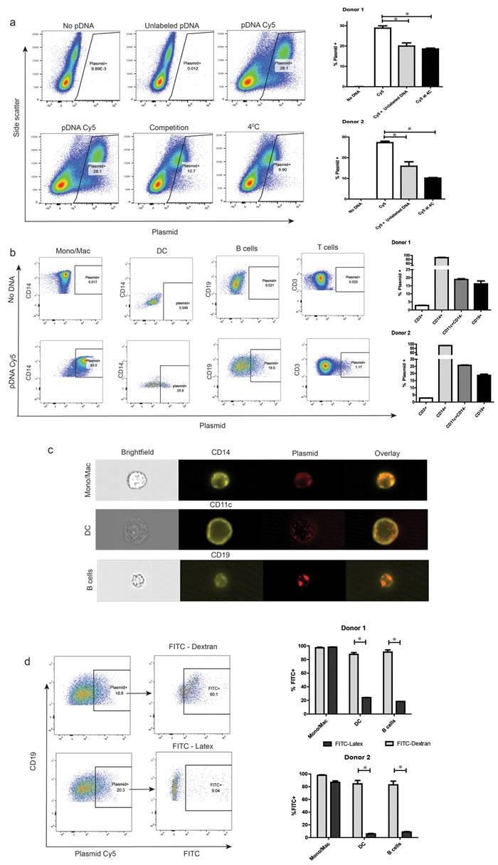
a. Autologous monocyte-derived DCs were added to cryopreserved human PBMCs. Cells were washed and re-suspended in PBS. Covalently labeled plasmid DNA was then added to samples that were either pre-incubated at 37°C or 4°C, both alone or along with 5μg/mL unlabeled DNA for 1hr, washed, and analyzed by flow cytometry. Dot plots of live single cells in each of the conditions are represented, with side-scatter and plasmid-associated fluorescence on the axes (left). Plasmid-associated fluorescence fractions in the different conditions for two representative donors (right). b. Cells were treated as above, and then stained for relevant surface markers before flow cytometric analysis. Subset-wise dot-plot representation of plasmid-associated fluorescence from a representative donor (left). Data on subset-wise association from two representative donors (right). c. Samples were treated as above, and analyzed for internalization of plasmid by imaging cytometry at a 60X magnification. Shown is a representative cell image of each APC cell type with internalized plasmid. d. Cells were treated as above, in addition to co-incubation with either FITC-Dextran or FITC-Latex beads. Plasmid+ APC sub-populations were then assayed for their association with either reagent. Representative dot plots showing extent of pinocytosis (FITC-Dextran+, left-top) and phagocytosis (FITC-Latex+, let-bottom) in plasmid positive B cells. The right panel is a graphical representation of data from similar studies utilizing cells from 2 separate donors. For all panels, * denotes a p-value <0.05, two-sided t-test. Data is representative of at least 5 independent experiments, involving at least 5 separate donors. Error bars represent mean + SEM.