
Figure 5. US28 signaling results in increased cell proliferation and metabolic reprogramming.
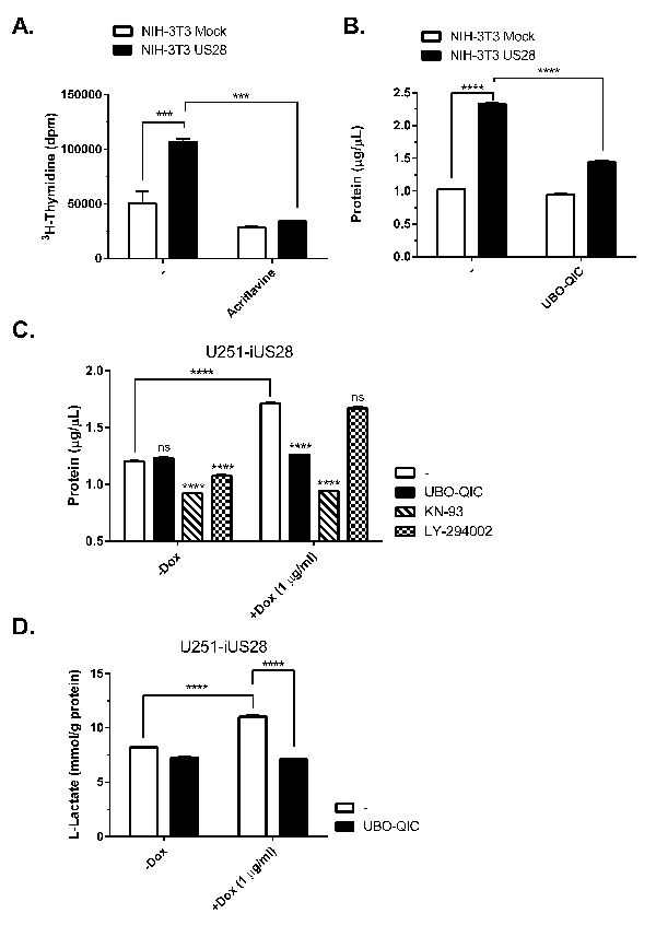
A. Stably transfected NIH-3T3 cells with US28 or empty vector were treated with acriflavine for 48 hours. Proliferation of the cells was determined by 3H- thymidine incorporation assay. B. Mock and US28 NIH-3T3 cells were synchronized and subsequently treated with UBO-QIC for 48 hours before lysates were prepared and protein content was determined using a BCA assay C., D. Synchronized U251 cells with inducible US28 expression were cultured for 48 hours with/without doxycycline and pharmacological inhibitors. (D) The lysate protein content was determined using a BCA assay, E. whereas cell culture medium supernatant was analyzed for L-Lactate levels using a cell-based glycolysis assay. The L-Lactate levels are normalized to the total protein content. Significant differences between conditions are depicted by asterisks (***= P < 0.001, **** = P < 0.0001).