
Figure 6. US28 contributes to HCMV-dependent HIF-1 activation in U251 glioblastoma and HFF cells.
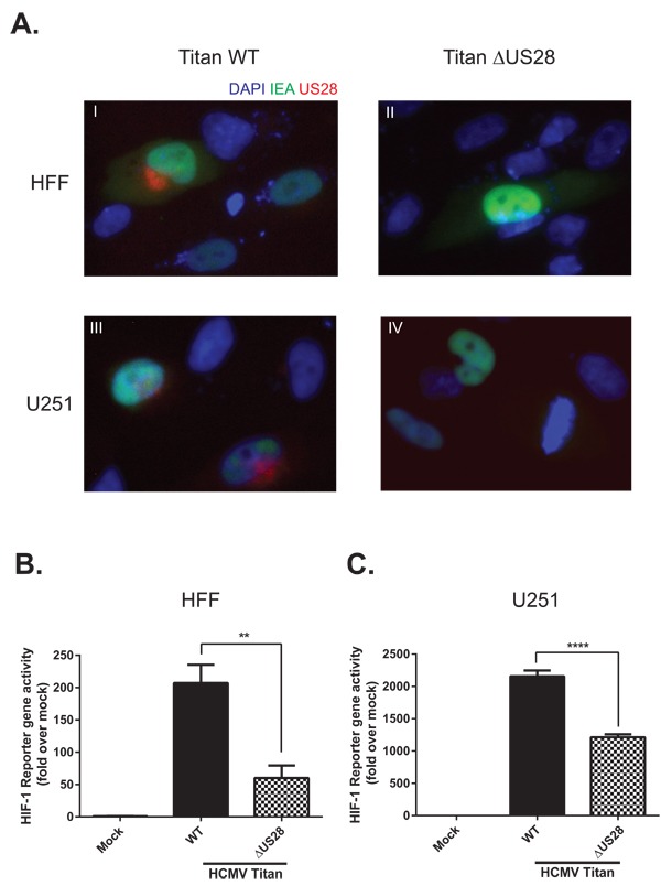
A. Immunofluorescence of HCMV Titan WT and ΔUS28 infected U251 or HFF cells 48 hours post-infection at MOI2. Cells were stained with US28 antibody (red fluorescence), anti-immediate early antigen (IEA) antibody (green fluorescence) and DAPI (blue fluorescence). Additionally, since a GFP tag is incorporated in the viral genome, the cytosol of infected cells can also be detected with green fluorescence. Panel I-IV display IEA-specific staining in the nuclei and GFP in the cytosol of HCMV Titan-infected cells. Panels I and II display US28-specific staining in primarily the perinuclear region of Titan WT infected cells, which cannot be detected in cells infected with the US28 deletion mutant (ΔUS28). B. HFF or C. U251 cells were infected with HCMV Titan WT or ΔUS28 at MOI of 2. Mock and infected cells were transfected with the HRE reporter gene 24 hours post-infection, followed by HIF-1 activity measurement 48 hours post-infection. Significant differences between conditions are depicted by asterisks (** = P < 0.01, **** = P < 0.0001).