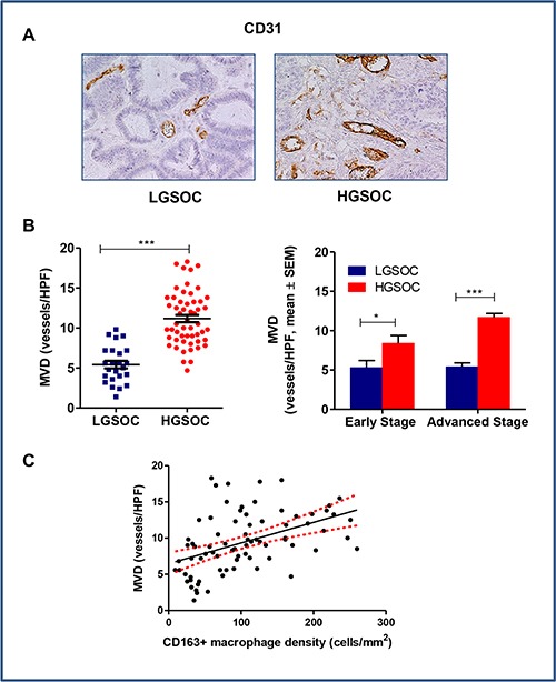
Figure 3. Tumor-associated angiogenesis in LGSOC and HGSOC tissue specimens.

(A) Representative pictures for immunohistochemical staining of CD31 in clinical samples of LGSOC and HGSOC. Magnification 20×. (B) Scatter plot shows MVD (Microvessel Density, vessels/HPF) values and mean ± SEM for the entire set of patients (see legend to Figure 2 for sample sizes, ***p < 0.0001). Bar graphs depict data (mean ± SEM) following stratification per stage (see legend to Figure 2 for sample sizes, *p = 0.04, ***p < 0.0001). (C) The Spearman rank correlation showed a significant positive correlation between MVD and CD163+ macrophages density (cells/mm2) (n = 80, p < 0.0001).
