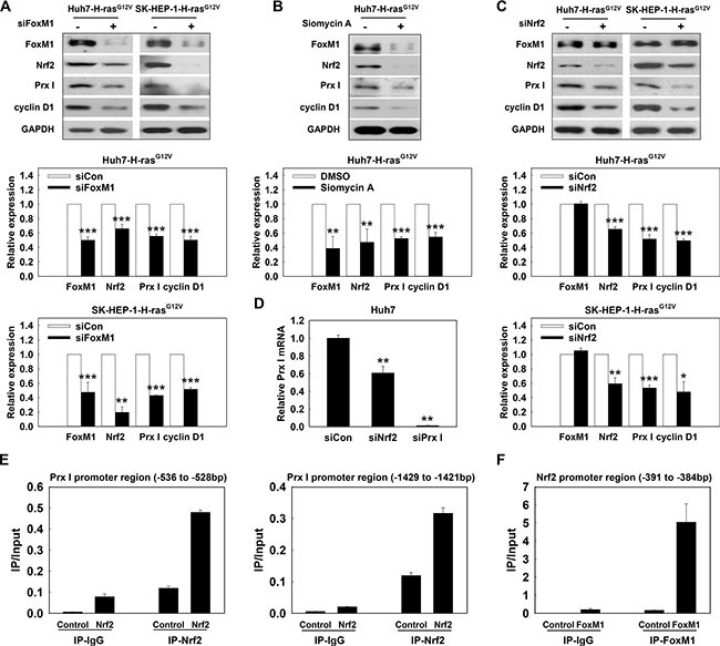
Figure 6. Oncogene Ras activated the ERK/FoxM1/Nrf2/Prx I pathway.

(A) Huh7-H-rasG12V cells and SK-HEP-1-H-rasG12V cells were transfected with siFoxM1 for 72 h; (B) Huh7-H-rasG12V cells were treated with Siomycin A (10 μM) for 24 h. After incubation, the cell lysates were used to determine protein expression. (C) After HCC-H-rasG12V cells transfected with siNrf2 for 72 h, the protein expression of FoxM1, Nrf2, Prx I, cyclin D1, and GAPDH were detected in the cell lysates. In A and C, *p < 0.05, **p < 0.01, ***p < 0.001 compared to siCon. In B, **p < 0.01, ***p < 0.001 compared to DMSO. (D) Prx I mRNA level was detected in Huh7 cells transfected with siRNA (scramble, Nrf2 or Prx I) for 48 h, **p < 0.01 compared to siCon. (E and F) CHIP assay was performed to detect the Nrf2 binding sites in the Prx I promoter region (–1429 to –1421), (–536 to –528) (E) or FoxM1 target site in the Nrf2 promoter region (–391 to –384) (F). HCC-H-rasG12V cells; Huh7- H-rasG12V and SK-HEP-1- H-rasG12V cells. The data were repeated in at least three separate experiments.
