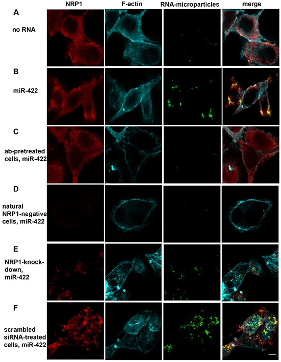
Figure 2. Biotinylated miR-422 conjugated to fluorescent streptavidin-coated microparticles binds to NRP1 expressed by ACHN cells and translocates into the cytoplasm.

The uptake of the beads is visualized by confocal microscopy. The fluorescent beads (green) are found in z- sections cutting through the nucleus. They co-localize with NRP1 (red; see also Figure 3). Cells are counter-stained for f-actin (cyan). A. Spontaneous uptake of the unconjugated beads is very low. B. Unassisted uptake of miR-422 conjugated to the beads by ACHN cells. C, D. Cells pre-treated with a blocking anti-NRP1 antibody (ab), or natural NRP1-negative cells, uptake negligible amount of miR-422-bead conjugate. E, F. Similarly, knockdown of NRP1 by siRNA severely depressed bead uptake, as compared to the scrambled siRNA control. Cells were transfected with siRNA using the unassisted translocation protocol. The data is representative of 6 independent experiments.
