
Figure 5. Proliferation and migration of 768-O and ACHN cells is regulated by miRNA mimics.
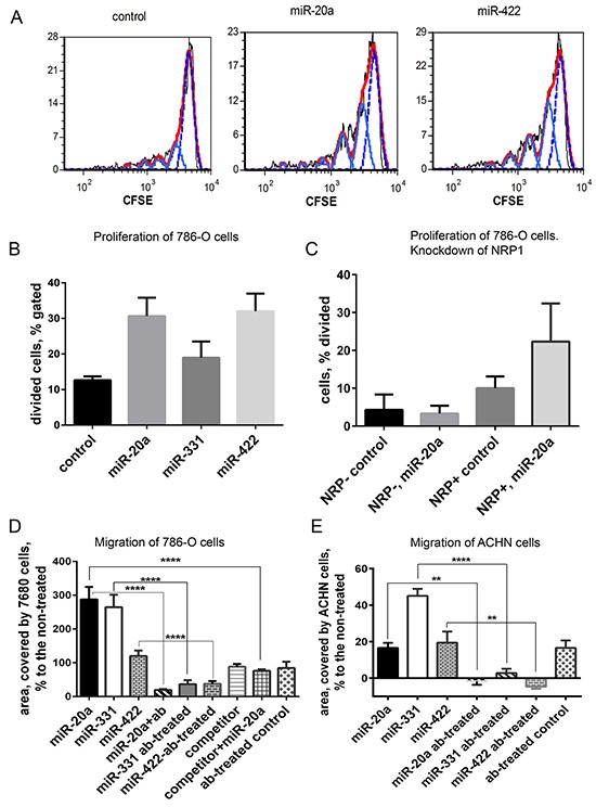
A. 768-O cells pre-loaded with CFSE and treated with miR-20a or miR-422. Cell division was traced by flow cytometry 24 h later. Generations of divided cells are depicted light blue, undivided cells are shown with dotted blue line, red is calculated fit line. B. Division of 768-O cells is activated by miR-20a, miR-331, and miR-422. C. Prior knockdown of NRP1 with siRNA abrogated the response of 786-O cells to miR-20a in the proliferation assay. D. Migration of 768-O cells pre-treated or not with anti-NRP1 antibody (ab) and treated with miR-20a, miR-331, miR-422, or competitor RNA in the wound-scratch assay. In case of the miRNA competition the cells were pre-treated with the competitor 30 min before adding the equal amount of miR-20a. The data is acquired after 20 h and presented as relative area covered by the migrating cells: Ar=100×(Atreated-Anon-treated)/Anon-treated. E. Migration of ACHN cells treated as in D. The data is representative of three independent experiments. The data (panels B-E) is presented as Mean±SEM, and statistical significance is denoted as: * P<0.05; ** P<0.01; **** P<0.0001.