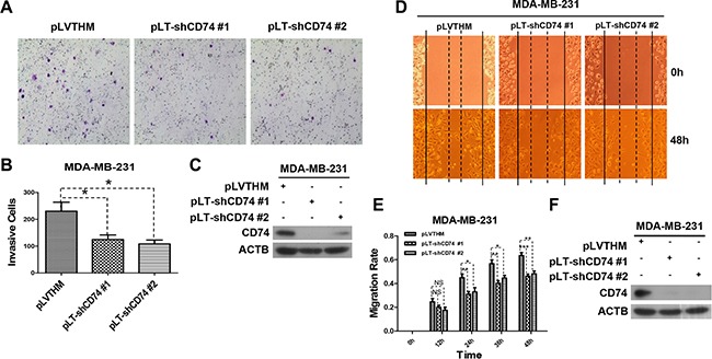
Figure 2. CD74 knockdown inhibited cell migration.

(A) MDA-MB-231 cells were transfected with pLVTHM (control) or pLT-shCD74 vectors (pLT-shCD74 #1 and pLT-shCD74 #2) for 48 h. Then, the cells were resuspended and counted, and 4 × 10⁵ cells in 500 μl of serum-free DMEM 3.7 medium were added to the upper well, while the lower well contained NIH3T3 conditioned medium (600 μl). The cells were incubated in a humidified culture incubator for 48 h. Representative images of cell invasion are shown. (B) The invading cells were counted. The error bars represent the SD, *P < 0.05. (C) MDA-MB-231 cells in each group were lysed after transfection, and the CD74 protein level was detected by western blotting. (D) MDA-MB-231 cells were transfected with pLVTHM or pLT-shCD74 plasmids, and a wound-healing scratch assay was conducted when the cells had grown to confluence. The images represent experiments at 0 and 48 h for each group. (E) The average width was recorded every 12 h, and differences between the treatment groups were analyzed by t-test. The error bars represent the SD, *P < 0.05; **P < 0.01; ***P < 0.001; ns, not significant. (F) Cells in different groups were collected and lysed at the end of a wound-healing scratch assay, and CD74 downregulation was detected by western blot.
