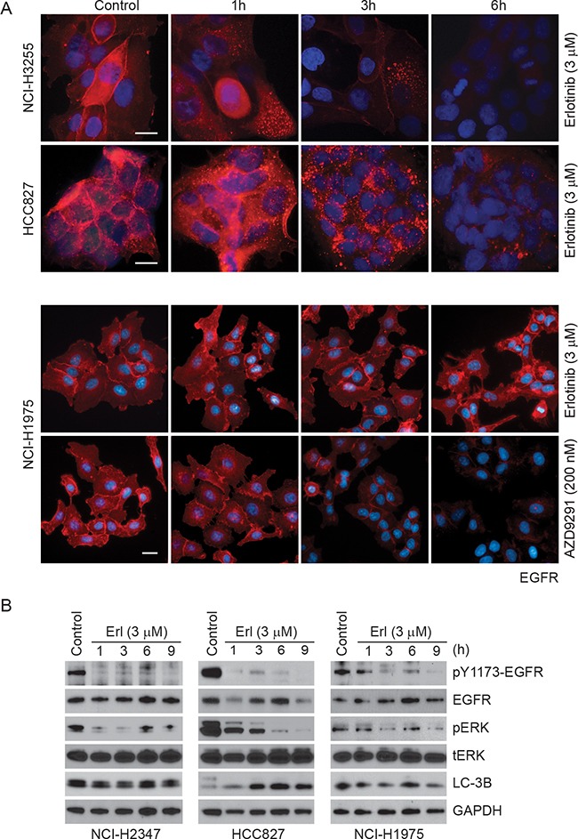
Figure 5. Time-dependent changes in EGFR immunofluorescence upon erlotinib/AZD9291 treatments in NSCLC cells.

A. HCC827 (delE746-A750), NCI-H3255 (L858R) and NCI-H1975 (L858R/T790M) cells were treated either with DMSO (control) or with 3 μM erlotinib/200 nM AZD9291 for the indicated time points. Cells were fixed and stained with EGFR antibody (red) and DAPI (blue, DNA dye) as described in the Materials and Methods. Representative images showing decreased EGFR immunofluorescence in erlotinib-sensitive HCC827 and NCI-H3255 cells. AZD9291 also reduced EGFR immunofluorescence even in NCI-H1975 cells. Scale bars, 10 μm. B. NCI-H2347, HCC827, and NCI-H1975 cells were either treated with DMSO (control) or with 3 μM erlotinib for the indicated time periods. Cell lysates were subjected to immunoblotting using the indicated antibodies.
