
Figure 5. Expression level of LASP1 inversely correlates with miR-145 level in vitro, in vivo and in CRC patients' tissue specimens.
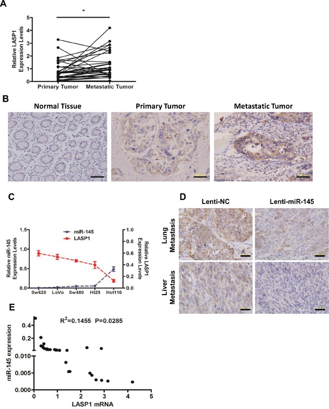
A, B. Expression levels of LASP1 in matched (normal mucosa) primary tumor and metastatic tumor tissues from 33 CRC patients as detected by qRT-PCR analyses (A) and IHC staining (B, from No. 5 of the 33 patients). * P < 0.05 (Paired t-test). Scale bars are 100 μm. C. Expression levels of miR-145 and LASP1 in Hct116, Ht29, LoVo, Sw480 and Sw620 cells were detected by qRT-PCR. Data represent the mean ± s.e.m. of three independent experiments. D. Representative photographs of anti-LASP1 IHC staining of lung (upper) or liver (lower) metastatic tumor tissues from Sw620/Lenti-NC and Sw620/Lenti-miR-145 nude mice groups. Scale bars are 50 μm. E. The correlation of LASP1 mRNA and miR-145 in metastatic tumor tissues from 33 CRC patients. The Pearson product-moment correlation coefficient and significance level are indicated.