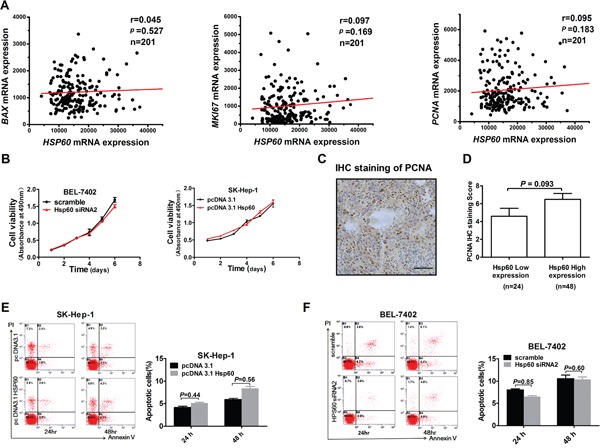
Figure 6. Hsp60 has no effect on proliferation and apoptosis of HCC cells.

A. Scatter plot analysis of correlation between mRNA expression levels of Hsp60 and BAX, KI67 and PCNA based on TCGA. Correlation coefficients (r) and levels of statistical significance (P value) were shown as indicated. B. MTS assay for the assessment of HCC proliferation ability. BEL-7402 and SK-Hep-1 cells were transiently transfected with Hsp60 siRNA and Hsp60 expression vector, respectively. Then cells were re-seeded for MTS cell viability assay 24 h after transfection. Absorbance at 490nm was measured at 1, 2, 3, 4, 5 and 6 days after reseeded. Values shown are the mean ± SD from at least three independent experiments. C. Representative IHC staining images of PCNA from HCC patient (Scale bars, 100μm). D. PCNA IHC score in HCC with low and high HSP60 expression. No statistically significant difference of PCNA expression level was observed between groups with high and low Hsp60 IHC score. E, F. Apoptosis analysis by flow cytometry in SK-Hep-1 and BEL-7402 cells treated as indicated by staining with Annexin V-FITC and propidium iodide (PI).
