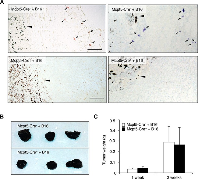
Figure 1. Mast cells do not influence the growth of subcutaneous melanoma tumors.

Melanoma cells (B16.F10) were injected subcutaneously into the flanks of either Mcpt5-Cre− or Mcpt5-Cre+ mice. After 1 or 2 weeks, tumors were excised. A. Staining of subcutaneous tissue of Mcpt5-Cre− or Mcpt5-Cre+ mice with either chloroacetate esterase (left panels) or toluidine blue (right panels). Note the presence of chloroacetate esterase- and toluidine blue-positive mast cells (marked with arrows) in tissue from Mcpt5-Cre− animals (upper panels) and the absence of mast cells in tissue from Mcpt5-Cre+ mice (lower panels). Note also that mast cells are abundant at a distance of ∼300 μm from the tumor border (arrowhead), but not detectable within the tumor mass. Original magnification = 200 x; Bar = 100 μm. B. Representative images of tumors excised from Mcpt5-Cre− and Mcpt5-Cre+ mice 2 weeks after injection of 0.5 − 106 B16.F10 cells. Bar = 5 mm C. Tumor end weights 1 or 2 weeks after injection of 0.5 − 106 B16.F10 cells. Results are shown as mean ± SD; n = 4-7.
