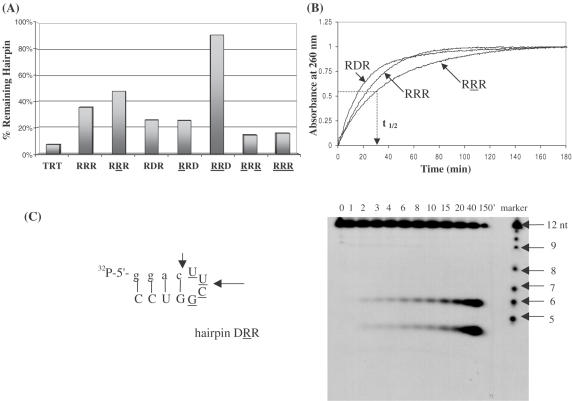
Figure 8.
(A) Biological stability of hairpin aptamers after treatment with 0.5× reticulocyte lysate for 18 h at 37°C. The 2′,5′-RNA loop enhances hairpin biostability. TRT does not form any hairpin structure and serves as a control. Equal amounts of 5′-[32P]-labeled oligomers were prepared in separate tubes containing 60 mM Tris (pH 7.8), 60 mM KCl, 5 mM MgCl2 and 5 mM K2HPO4. After addition of rabbit reticulocytes lysate, the reaction mixtures were incubated at 37°C for 18 h. Analysis was done on 16% denaturing polyacrylamide gels. (B) Plots of absorbance (at 260 nm) versus time of exposure of hairpins to nuclease P1. The time necessary to cause 50% degradation of the hairpin structure (t1/2) was calculated at absorbance = 0.5. (C) Gel degradation assay showing the specific cuts induced by nuclease P1 in hairpin DRR. The degradation ladder is formed via alkaline hydrolysis of the hairpin.

