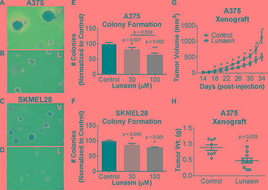
Figure 1. In vitro efficacy of lunasin in malignant melanomas.

Representative images of colonies grown in soft agar for vehicle-treated (A, C) and Lunasin-treated (B, D) A375 (top panels) and SKMEL-28 (bottom panels) cells (magnification at 40×). Scale bars on images represent 100 μm. Anchorage-independent growth conditions sensitized melanoma cells to Lunasin resulting in a significant decrease in colony formation in A375 (E) and SKMEL-28 (F) cells. Statistical significance between treatment groups is denoted by a different number of asterisks (*, **) and p-values are provided for each significant difference. Error bars on graphs represent mean ± S.D. For xenograft studies (2.5 × 10⁶ A375 cells were injected s.c. into nude mice and subsequently treated with vehicle (n = 8) or Lunasin (n = 10) for a total of 34 d. Lunasin reduced tumor volume by 55% (G) and wet tumor weight by 46% (H). Lunasin-treated mice differed significantly in tumor volume (p < 0.001) from control treated mice. The corresponding reduction in wet tumor weights were determined to be significant by unpaired student's t-test (p = 0.003) and denoted by an asterisk (*). Error bars represent mean ± S.E.M.
