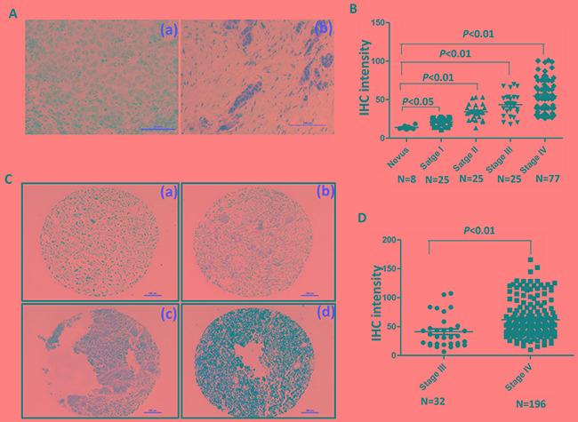
Figure 7. FOXC1 expression is associated with progress of melanoma.

A. Representative IHC pictures of FOXC1 in melanoma tissues. (a) Negative control. (b) FOXC1 expression. B. FOXC1 expression was increased as progress of melanoma. FOXC1 expression was significantly lower in nevus than cutaneous melanoma at different stages. C. Representative IHC photographs of FOXC1 expression in TMA (Stage III N=32; Stage IV=196). (a) Negative. (b) Weak. (c) Middle. (d) Strong. D. There is higher FOXC1 expression in TMA of stage IV (N=196) of than that in TMA of stage III (N=32). Error bars, s.d. (*p <0.05, **p<0.01).
