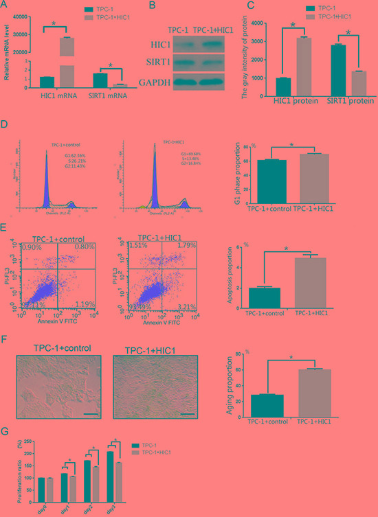
Figure 5. Effects of overexpression of pcDNA3.1(+)-flag-HIC1 on TPC-1 cells.

(A) qRT-PCR analysis of HIC1 and SIRT1 mRNA expression levels in TPC-1 cells after transfection with an HIC1 overexpression plasmid. (B–C) HIC1 and SIRT protein expression levels were determined in TPC-1 cells by Western blot (n = 3). GAPDH was used as an internal control. Flow cytometry was used to confirm that TPC-1 cells transfected with pcDNA3.1(+)-flag-HIC1 were arrested in the G1 phase (D) and that apoptosis was induced (E). (F) β-galactosidase staining showed that transfection with pcDNA3.1(+)-flag-HIC1 induced cellular senescence. Scale bar = 100 μm. (G) Growth of TPC-1 transfected with pcDNA3.1(+)-flag-HIC1 was inhibited. *p < 0.05 compared to the TPC-1 group transfected with control plasmid using a Student's t test.
