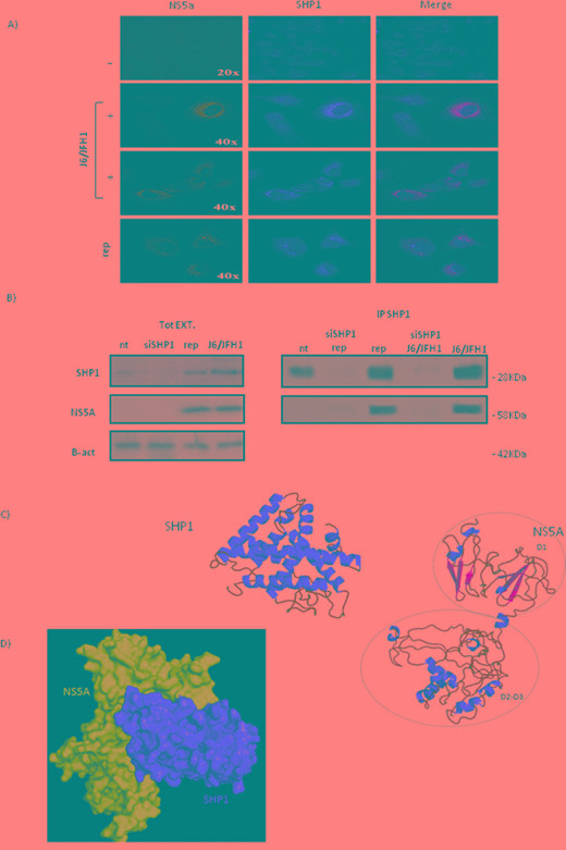
Figure 3. Interaction between SHP1 and NS5A.

A. Confocal double immunofluorescence microscopy of: NS5A and endogenous SHP1 on Huh7.5 (first horizontal panel), Huh7.5 infected J6/JFH1 infected cells (second and third panel), and replicon system (fourth horizontal panel), 72 hours post infection. Merge of the two analyzed proteins is on the right. B. SHP1 and NS5A coimmuno-precipitation assay, in Huh7.5 J6/JFH1 cells and JFH1 A4 replicon systems. On the left, WB of total protein extracts; on the right, immuno-precipitation in Huh7.5 J6/JFH1 cells and JFH1 A4 replicon systems before and after si-SHP1. Data are representative of three independent experiments. C. Model of SHP1 and NS5A: alpha-helices and beta-sheets are depicted by red and yellow, respectively. D. Model of the complex between SHP1 (in red) and NS5A (in cyan) by surface representation.
