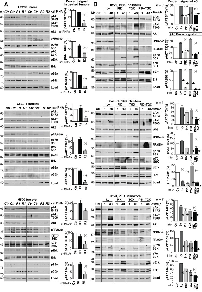
Figure 7. PIK3R2 depletion, but not PI3K inhibitors, induces stable PI3K pathway inhibition.

A. Tumor xenografts were obtained using H226, CaLu-1 and H520 lung SQCC cells expressing inducible PIK3R1 or PIK3R2 shRNA. When tumors reached ~50 mm3, mice were treated with doxycycline in drinking water (2 mg/ml); they were sacrificed when tumors began to diminish. Normalized tumor extracts were examined by WB with indicated antibodies. B. H226, CaLu-1 and H520 cells were cultured in exponential growth were incubated with: vehicle (DMSO, 1:103 V:V), Ly294002 (5 μM), PIK75 (200 nM), TGX221 (30 μM), or PIK75 (200 nM) plus TGX221 (30 μM) for the last 1 h of culture or during 48 h; extracts were tested in WB. Mr indicates relative mobility. In both A and B, the pAkt or pPRAS40 signal was measured and normalized to the loading control (Akt or tubulin, respectively), and compared to that in controls (DMSO, considered 100%). Graphs show percentage of signal compared to maximal as mean ± SD at 48h of treatment. Arrows on the right side of bars show the percentage of signal compared to maximal detected after 1h of treatment.* P <0.05; **P <0.01; unpaired Student's t test with Welch correction.
