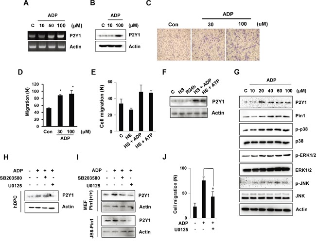
Figure 2. ADP-induced hDPC migration is involved in the MAPK pathway.

A, B. The mRNA and protein expression of the ADP-induced P2Y1 receptor in hDPCs was determined by RT-PCR and Western blot analyses. The cells were treated with ADP at the indicated concentrations for 24 h. C, D. In vitro migration assays. The cells were treated with ADP (30 or 100 μM) for 24 h, and the migration assays were performed in 12-well plates. After 24 h, hDPCs were fixed, stained with Mayer's hematoxylin, mounted, imaged and counted at 100X magnification using an optical microscope to assess the extent of migration. *P < 0.05 was considered significantly different from the control. E, F. Effects of ADP and ATP on the migration of and P2Y1 expression in cells subjected to thermal stress. The cells were exposed to 42°C for 30 min and incubated with or without 100 μM ADP or ATP for 24 h. The graphs show the quantitative evaluation of the migration rates. The levels of P2Y1 protein were detected by Western blot analysis. G. ADP stimulation of MAPK. The ERK1/2, JNK, and p38 phosphorylation levels in hDPCs stimulated with ADP at the indicated concentrations for 24 h were determined through Western blot analysis. H, I. Effects of MAPK inhibitors on ADP stimulation of MAPK pathways and cell migration. The phosphorylation levels of ERK1/2 and p38 in hDPCs stimulated with ADP (100 μM) for 24 h in the absence (control) or presence of 20 μM U0126 or SB203580 (SB) were determined by Western blot analysis. J. Effects of U0126 (20 μM) on ADP-stimulated hDPC migration. *P < 0.05.
