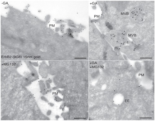
Figure 4. Ultrastructural localization of ERBB2 in SKBR3 cells treated with GA and proteasome inhibitors.

Cryosections of SKBR3 cells were labeled with antibodies against ERBB2 (9G6), followed by protein A-coated (15nm) colloidal gold (black dots). The labeling shows that ERBB2 localized to the highly ruffled plasma membrane in untreated cells A., and mainly in multivescicular bodies (MVBs) in GA-treated cells B. Cells treated with 10μM MG132 show plasma membrane localization of ERBB2, as in untreated cells C. Cells co-treated with GA and MG132 show intracellular localization of ERBB2 in morphologically resembling early/recycling endosomes (D).
