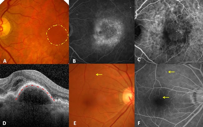
Figure 1.

Illustration of diabetic choroidal microvascular proliferation. A. Fundus photography demonstrating sporadic retinal microaneurysms and a submacular round dark reflection involving the fovea (yellow circle), without drusen. B. Late stage of fluorescein angiography showing macular leakage, multiple hyperfluorescent dots, and the normal background fluorescence originating from RPE except for leakage. C. Late stage of indocyanine green angiography indicating multiple hyperfluorescent dots and CNV. D. Foveal horizontal optic coherence tomography showing subfoveal fluid together with RPE detachment (red line). E. Fundus photography of the right eye only demonstrated macular retinal microaneurysms. F. Late-stage fluorescein angiography of the right eye revealed more retinal microaneurysms and normal background fluorescence originating from the RPE.
