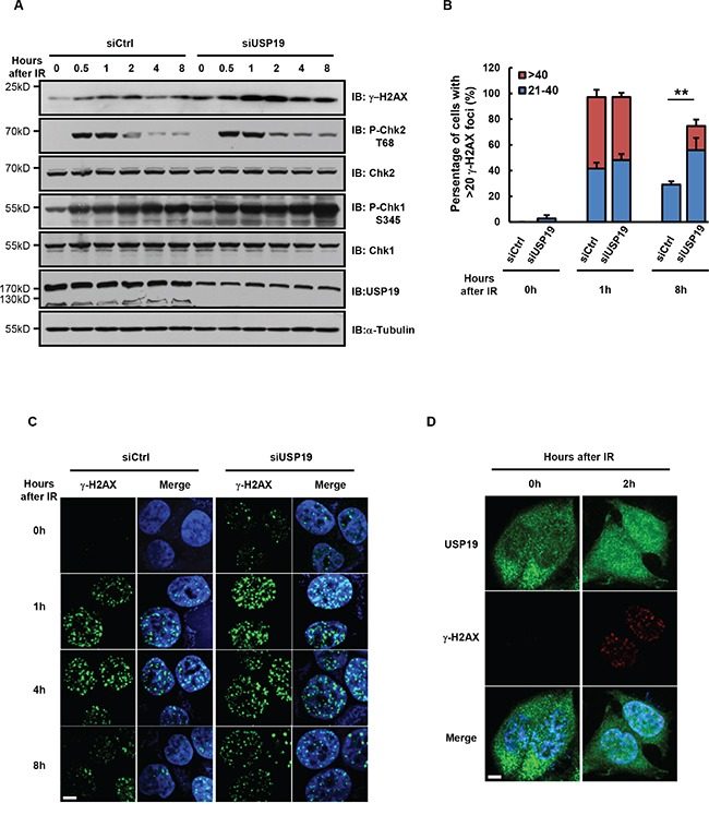
Figure 2. Cells depleted with USP19 show accumulation of DNA-damage.

A-C. HCT116 were transfected with control or USP19 siRNA and irradiated with 5Gy γ-ray. At various time points after irradiation, (A) immunoblot analysis were performed to detect the level of γ-H2AX, phosphorylation of Chk1(P-Chk1 S345), phosphorylation of Chk2(P-Chk2 T68), Chk1, Chk2 and USP19; (B) Irradiated cells were immunostaining with γ-H2AX antibody (green) and DAPI (blue), fraction of cells with the noted numbers of γ-H2AX foci per cell were quantified, data are shown as mean ± S.D; *p<0.05, **p<0.01 (C) representative images of γ-H2AX foci. Scale bar, 5 μm. D. USP19 localization after irradiation. Control cells and USP19 knockdown cells were irradiated with 5Gy γ-ray, follow by immunostaining with USP19 antibody (green), γ-H2AX antibody (red) and DAPI (blue). Scale bar, 5 μm.
