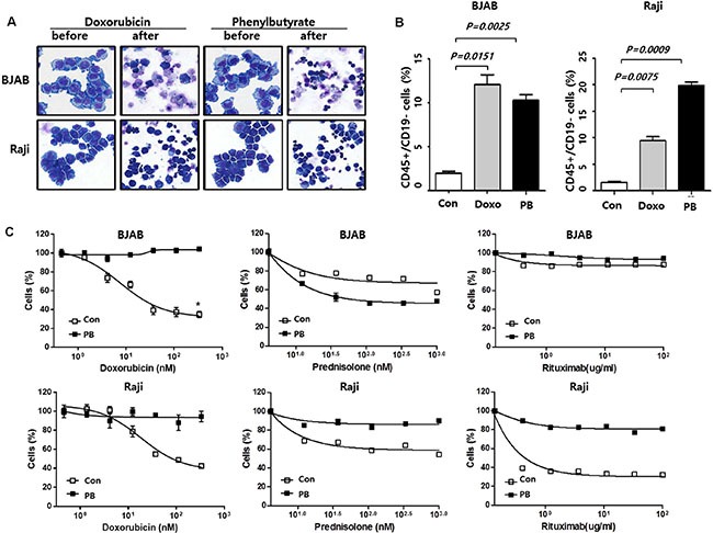
Figure 1. Generation of B-cell lymphoma cells surviving drug treatment.

(A) Morphology of BJAB and Raji cells after 48 h incubation with doxorubicin (300 nM) or phenylbutyrate (8 mM): Original magnification, x 400; May–Grünwald–Giemsa staining. (B) Flow cytometry analysis of the CD45+/CD19− cell population and comparison of CD45+/CD19− cell fraction among control cells (con), doxorubicin (Doxo) and phenylbutyrate (PB)-treated surviving cells. (C) Dose-response curves shows higher viability of phenylbutyrate (PB)-treated surviving BJAB and Raji cells than control cells (con) when cells are seeded at a density of 5 × 10⁴ cells per well in 24-well plates, treated with the indicated doses of doxorubicin, prednisolone and rituximab. Data represents means ± SEM of three independent experiments.
