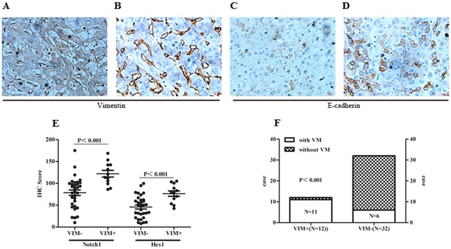
Figure 7. Notch1 associated with EMT related biomarkers in HCC tissues.

A. Positive Vimentin staining in tumor cells and microvascular wall. B. Positive vimentin staining only in microvascular wall. C. Expression of E-cadherin in HCC specimens with positive Vimentin staining in tumor cells. D. Expression of E-cadherin staining in HCC specimens with only positive Vimentin staining in microvascular wall. Original magnification 400×. E. Expression of Notch1 and Hes1 were elevated in those specimens with positive Vimentin staining in tumor cells (Notch1: 121.99±26.13 vs. 78.63±35.88; Hes1: 76.49±21.18 vs. 45.93±25.48). F. Distribution of cases with positive Vimentin staining in specimens with and without VM. VIM+: Positive Vimentin staining in tumor cells; VIM-: Negative Vimentin staining in tumor cells.
