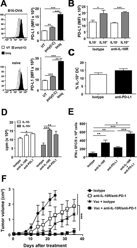
Figure 6. Blockade of vaccination-induced IL-10/PD-L1 in DC potentiates T-cell responses and increases antitumor therapeutic efficacy.

(A) Naive or B16-OVA tumor-bearing Vert-X mice (n = 4/group) were vaccinated with OVA+Imiquimod, OVA+poly(I:C) or left untreated. Two days later PD-L1 expression was determined in DC. (B) Vert-X mice (n = 4/group) were vaccinated with OVA+Imiquimod with or without IL-10 blockade and PD-L1 expression was determined two days later in IL-10− and IL-10+ DC. (C) Vert-X mice (n = 4/group) were vaccinated with OVA+Imiquimod with or without PD-L1 blockade and the proportion of IL-10+ DC was determined two days later. (D) IL-10− and IL-10+ DC obtained from OVA+Imiquimod-vaccinated mice were used to stimulate OT-I CD8 T-cells in the presence of OVA(257–264) plus control or IL-10R- or PD-L1-blocking antibodies. T-cell proliferation was determined three days later. (E) C57BL/6 mice (n = 4/group) were vaccinated with OVA+Imiquimod together with control or IL-10R- or PD-L1-blocking antibodies and one week later OVA(257–264)-specific responses were determined by ELISPOT. (F) C57BL/6 mice (n = 7–8/group) bearing 5 mm B16-OVA tumors were treated with three weekly vaccination cycles with OVA+Imiquimod with or without IL-10R/PD-1-blocking antibodies and tumor volume was monitored. Results are representative of 2–3 independent experiments.
