
Figure 2. The 3A2 Fab antibody inhibits the functional activity of cellular MT1-MMP.
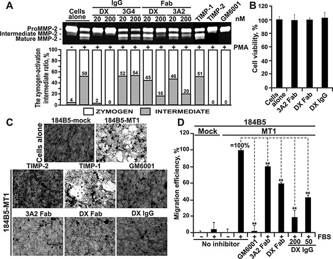
A. The 3A2 Fab and the DX2400 Fab and IgG antibodies inhibited activation of the proMMP-2 zymogen by cellular MT1-MMP in HT1080 cells. Top, to induce proMMP-2 activation, cells were stimulated using phorbol 12-myristate 13-acetate (PMA; 50 ng/ml). Cells were then co-incubated with the 3A2 and DX2400 antibodies (20-200 nM, each) and also with the non-inhibitory MT1-MMP 3G4 IgG antibody (20-200 nM), TIMP-1 (1,000 nM), TIMP-2 (100 nM) and GM6001 (1,000 nM) controls. Medium aliquots were next analyzed by gelatin zymography to identify the status of MMP-2. Cells alone, no inhibitors were added to the cells. Bottom, the digitized zymogen:activation intermediate ratio in the MMP-2 samples. White and grey rectangles, zymogen and activation intermediate, respectively. The numbers indicate the percentage of the activation intermediate relative to the total combined amount of the zymogen and the intermediate. B. The 3A2 Fab and the DX2400 Fab and IgG do not affect cell viability. Normal mammary epithelial 184B5 cells were incubated alone (cells alone) or co-incubated with the antibodies (1,000 nM, each). Cell viability was measured using a luminescent ATP-Lite assay. Data are means ± SE from three individual experiments performed in triplicate. C. The 3A2 Fab antibody inhibits COL-I degradation by cellular MT1-MMP. MT1-MMP-deficient 184B5-mock and MT1-MMP-overexpressing 184B5-MT1 cells were plated onto COL-I layers and then incubated alone or co-incubated for 5 days with the 3A2 Fab (200 nM), DX2400 Fab or IgG (200 nM and 100 nM, respectively), TIMP-1 (1,000 nM), TIMP-2 (100 nM) or GM6001 (1,000 nM). After the removal of cells, COL-I was stained with Coomassie. The representative images from three independent experiments performed in triplicate are shown. D. Cell invasion through COL-I. 184B5-mock (mock) and 184B5-MT1 (MT1) cells (1×10⁵, each) were allowed to migrate alone (no inhibitor) or in the presence of the 3A2 or DX2400 Fab fragments (500 nM, each) or the indicated concentrations DX2400 IgG. GM6001 (1,000 nM) and 10% FBS were used as a control and a chemoattractant, respectively. Migration efficiency was calculated relative to MT1 cells, no inhibitor and 10% FBS (=100%). Data are means ± SE from three individual experiments conducted in triplicate. **, P < 0.05. DX, DX2400.