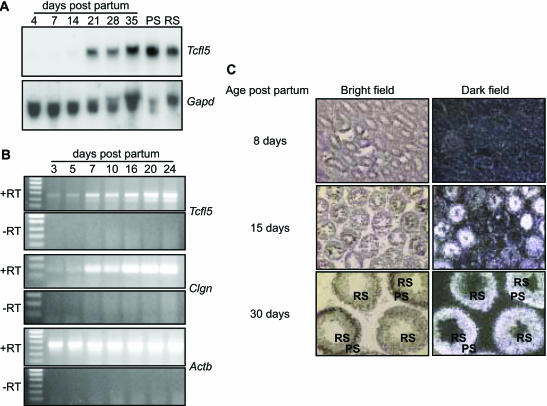
Figure 3.
Expression of Tcfl5 mRNA during spermatogenesis. (A) Northern-blot analysis of total RNA isolated from testis of mice of different ages (4-, 7-, 14-, 21-, 28- and 35-day-old mice) and purified spermatocytes (PS) and spermatids (RS). Only a faint signal is detected in day 4, 7 and 14 testis and the signal increases significantly between day 14 and day 21. Spermatocytes show a slightly higher expression level of Tcfl5 mRNA than spermatids. Loading of the different lanes was checked by hybridization with a Gapd cDNA. (B) Using RT–PCR, testicular RNAs from mice aged 3–24 days were analysed for Tcfl5 and Clgn expression. Both, the 455 bp Tcfl5 and the 166 bp Clgn PCR product intensified in later stages of spermatogenesis. Primers specific for the ubiquitously expressed Actb gene were used to show that equal amounts of material were used. The 448 bp Actb PCR product is not detected in the control experiment without reverse transcriptase (−RT), ruling out contamination with genomic DNA. (C) In situ hybridization on postnatal mouse testes from 8-, 15- and 30-day-old mice with an antisense Tcfl5 RNA probe shows that Tcfl5 mRNA expression starts in the spermatocytes. Little Tcfl5 mRNA signal is detected in the 8-day-old mouse testis. In the 15-day-old mouse testis, many tubule cross-sections show Tcfl5 mRNA signal, while other tubules show no signal. The cells that show the Tcfl5 mRNA signal are spermatocytes. Expression of Tcfl5 mRNA continues in spermatids as is shown in the 30-day-old mouse testis, where both spermatocytes and spermatids show the Tcfl5 mRNA signal. PS, pachytene spermatocytes; RS, round spermatids. With the sense probe no signal is visible, indicating the specificity of the antisense probe (data not shown).

