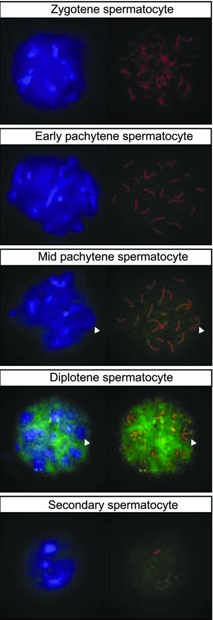
Figure 5.
Tcfl5 protein expression in nuclei of late spermatocytes. Using immunocytochemistry on spread nuclei of spermatocytes Tcfl5 protein is detected from mid-pachytene spermatocytes to secondary spermatocytes. Specificity of the antibody for Tcfl5 was verified by peptide competition assay (data not shown). Blue: DAPI DNA staining; green: P3-αTcfl5; red: αSycp3; and arrowheads indicate the XY body.

