
Figure 2. Effects of SAHA or/and MG132 on the cell cycle and apoptosis of glioma cells.
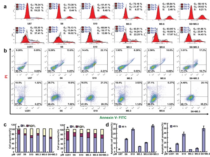
Flow cytometric analyses of glioma cell lines after PI staining showed that SAHA or/and MG132 treatment induced G2 arrest in a concentration-dependent manner in U87 and U251 cells after 48 h a, c. SAHA or/and MG132 exposure results in higher levels of apoptosis in U87and U251cells after 48 h b, d. Results are representative of 3 different experiments, and the data is expressed as mean ±standard deviation. Note: S6, SAHA 6 μM; M0.3, MG132 0.3 μM; S10, SAHA 10 μM; M0.6, MG132 0.6 μM; S6 + M0.3, SAHA 6 μM and MG132 0.3 μM. *p < 0.05, vs treatment groups.