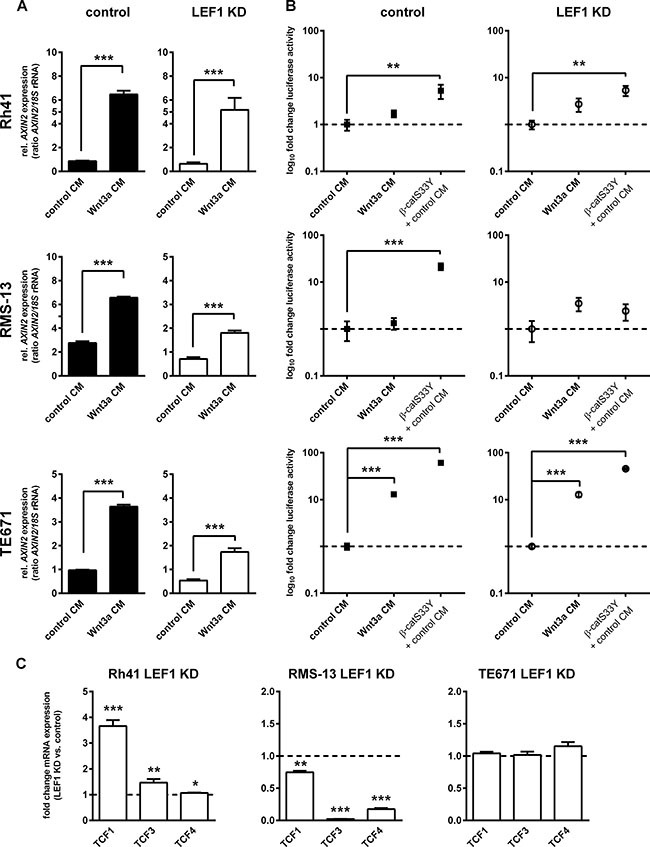
Figure 3. LEF1-dependent modulation of canonical WNT signaling activity in RMS cell lines.

(A) Expression of AXIN2 in Rh41 LEF1 KD, RMS-13 LEF1 KD and TE671 LEF1 KD and respective control cells in response to Wnt3a conditioned medium (Wnt3a CM) or control medium (control CM). Gene expression levels were normalized to 18S rRNA expression levels. Data represent mean+SEM of at least two independent experiments performed in duplicates and measured in triplicates. (B) To analyze β-catenin-dependent WNT signaling in response to LEF1 KD, cells were transfected with SuperTOPFlash (TOP) containing multiple TCF/LEF-binding sites and Renilla reporter plasmid for normalization. Luciferase activity was measured 5 days after transfection in response to Wnt3a or control CM. Transfection of the cells with pCl-neo-β-catS33Y (β-catS33Y) served as positive control. Data show the 95% confidence intervals of at least two independent experiments performed in duplicates and are depicted as fold luciferase activity to cells treated with control CM (set to 1; dashed line). (C) Expression of TCF1, 3 and 4 in Rh41 LEF1 KD, RMS-13 LEF1 KD and TE671 LEF1 KD are shown as fold expression to the respective control cells that were set to 1 (dashed line). Gene expression levels were normalized to GAPDH expression levels. Data represent mean + SEM of at least four independent experiments measured in triplicates. (A, B and C) *P < 0.05, **P < 0.01, ***P < 0.001 by Students t-test.
