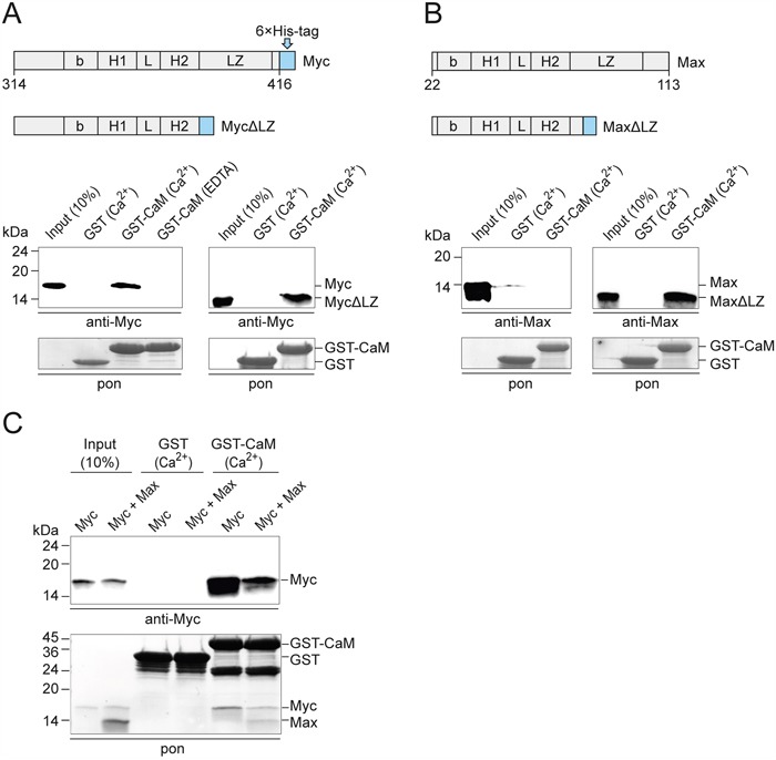
Figure 3. PPI of CaM and bHLH domains of recombinant Myc and Max.

A. GST pull-down experiments using recombinant v-Myc314-416 protein or v-Myc314-383 lacking the leucine zipper (MycΔLZ) and GST-CaM. Myc proteins were detected by immunoblotting using a Myc-specific antiserum. A section of the membrane with Ponceau S-stained GST bait proteins is shown (pon, lower panel). EDTA, 2 mM; Ca2+, CaCl2 0.5 mM. B. PPI analyses of recombinant Max22-113 protein or Max22-79 lacking the leucine zipper (MaxΔLZ) and GST-CaM. Max proteins were detected using a Max-specific antiserum. C. GST pull-down experiments using recombinant v-Myc314-416, Max22-113, and GST-CaM in the presence of 0.5 mM CaCl2. Where indicated, Myc and Max proteins were mixed using a threefold excess of Max protein. After SDS-PAGE and electroblotting, bait and prey proteins were visualized by Ponceau S-staining (pon, lower panel). Myc was detected by immunoblotting using a Myc-specific antiserum (upper panel).
