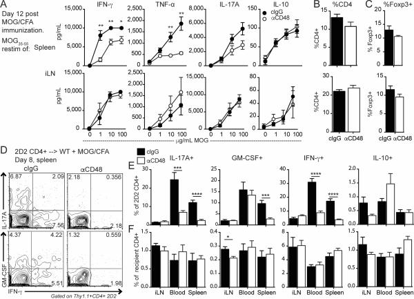
Figure 3. Anti-CD48 reduces antigen-specific cytokine production in the spleen but not the draining LN.
A-C WT mice were immunized with MOG35-55/CFA, and given anti-CD48 or cIgG 6 and 9 days later. Spleen and iLN were collected on day 12. A. Cytokines in supernatants from spleen (top) or iLN (bottom) cells restimulated with the indicated concentrations of MOG35-55 for 3 days. Percentages of CD4+ of total cells (B) and Foxp3+ of CD4+ (C) in the iLN and spleen ex vivo. D-F. WT mice were given 2D2 Thy1.1+ CD4+ T cells i.v., immunized with MOG35-55/CFA, and treated with anti-CD48 or cIgG 4 and 6 days later. Spleen, iLN and peripheral blood were collected on day 8 and restimulated with PMA/ionomycin. D. Representative intracellular cytokine staining in splenic 2D2 Thy1.1+ CD4+ T cells. E. Percentages of cytokine-positive cells among 2D2 cells in the spleen, iLN and blood, gated as in D. F. Percentages of cytokine-positive cells among recipient Thy1.1− CD4+ cells. Representative of 2 independent experiments with 5 mice per group (A-C), and 4 independent experiments with 3-7 mice per group (D-F).

