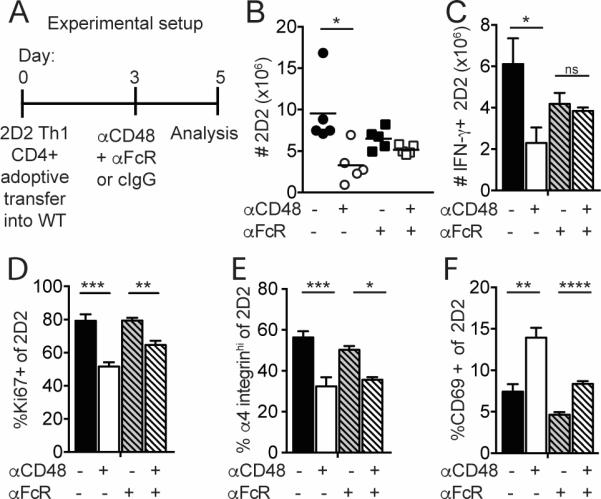
Figure 6. An FcγR blocking antibody limits the effect of anti-CD48 on 2D2 Th1 CD4+ Teff cells in vivo.

2D2 Th1 CD4+ Teff (Thy1.1+) cells were transferred i.v. into WT recipients that received anti-CD48 or cIgG plus anti-CD16/CD32 or isotype control on day 3. Spleen was collected on day 5 for analysis by flow cytometry as in Figure 5. A. Experimental setup. B. Numbers of 2D2 CD4+ in the spleen. C. Numbers of IFN-γ+ 2D2 cells after restimulation with PMA/ionomycin. Percentages of Ki67+ (D), α4-integrinhi (E) and CD69+ (F) of 2D2 CD4+ T cells. Representative of 4 independent experiments with 4-5 mice per group. Dots in B represent individual mice.
