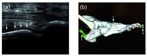
Figure 1. New imaging modalities for demonstrating serum urate deposition.
( A) Musculoskeletal ultrasound of a first metatarsal phalangeal joint (plantar longitudinal view) demonstrating a classic “double contour sign” (arrows), indicating the deposition of monosodium urate (MSU) crystals on the cartilage surface of the metatarsal head. ( B) Dual-energy computed tomography of a foot. Green areas indicate MSU deposition, and arrows indicate the presence of MSU deposition at the first distal interphalangeal joint, at the carpal metacarpal joint, and along the Achilles tendon.

