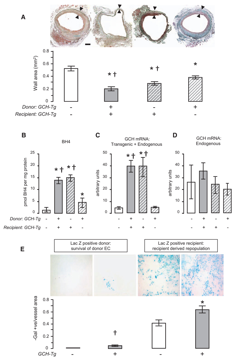
Figure 1. Endothelial overexpression of human GTP cyclohydrolase (GCH) reduces neointimal hyperplasia and enhances repopulation and survival of endothelial cell (EC) in vein grafts.
A, When vena cava was grafted into recipients of the same genotype, vein graft wall area at 28 days after surgery was significantly lower in GCH/apolipoprotein E (apoE)-knockout (KO; n=5) mice compared with apoE-KO mice (n=8). When vena cava of apoE-KO mice was grafted into GCH/apoE-KO recipients (n=6), the mean vessel wall area was similar to that seen when GCH/apoE-KO vena cava was grafted into GCH/apoE-KO recipients. When GCH/apoE-KO donor vena cava was grafted into apoE-KO recipients (n=8), there was a less marked, but still significant, reduction in lesion area compared with apoE-KO vena cava grafted into apoE-KO. Black bar represents 100 μm. One-way ANOVA overall P<0.0001. Bonferroni multiple comparison test *P<0.01 vs apoE-KO vein grafted into apoE-KO recipient and †P<0.05 vs GCH/apoE-KO vein grafted into apoE-KO. B, Tetrahydrobiopterin (BH4) levels in harvested vein grafts were significantly higher in GCH/apoE-KO compared with apoE-KO. When apoE-KO veins were grafted into GCH/apoE-KO mice, graft BH4 levels reached levels similar to the GCH/apoE-KO vein grafts. When GCH/apoE-KO veins were grafted into apoE-KO mice, BH4 levels were significantly higher than apoE-KO vein grafts grafted into matched recipients (n=5). One-way ANOVA overall P<0.0001. Bonferroni multiple comparison test *P<0.05 vs apoE-KO. †P<0.05 vs GCH/apoE-KO vein grafted into apoE-KO recipients. C, Vein graft mRNA levels of total GCH (transgenic+endogenous) were significantly higher in GCH/apoE-KO mice compared with apoE-KO. When apoE-KO veins were grafted into GCH/apoE-KO mice, mRNA levels were similar to the GCH/apoE-KO vein grafts. One-way ANOVA overall P<0.0001. Bonferroni multiple comparison test *P<0.001 vs apoE-KO. †P<0.001 vs GCH/apoE-KO vein grafted into apoE-KO recipients (n=4). D, Vein graft endogenous mouse GCH mRNA levels were not different in GCH/apoE-KO mice compared with apoE-KO, regardless of the source of the vein (n=4). E, When apoE-KO veins were grafted into apoE-KO/LacZ recipients, the presence of β-galactosidase (β-Gal)–positive staining (blue) on the surface of vein grafts at 28 days indicated recipient-derived EC repopulation (n=5). When GCH/apoE-KO veins were grafted into GCH/apoE-KO/LacZ recipients (n=6), EC coverage was increased compared with grafting apoE-KO veins into apoE-KO/LacZ recipients, indicating enhanced recipient-derived EC repopulation in GCH animals. Investigating EC survival, no β-Gal staining in apoE-KO/LacZ veins grafted into apoE-KO recipients could be detected. However, when GCH/apoE-KO/LacZ veins were grafted into GCH/apoE-KO recipients (n=5), a small proportion of β-Gal–positive cells remained evident on the luminal surface of the vein graft at 28 days, indicating enhanced EC survival from GCH transgenic donors. No β-Gal–positive cells were visualized in apoE-KO/LacZ veins grafted into apoE-KO recipients (n=5). Arrowhead denotes β-Gal–positive cell. Unpaired t test *P=0.02 vs apoE-KO/LacZ recipient. †P=0.02 vs apoE-KO recipient.

