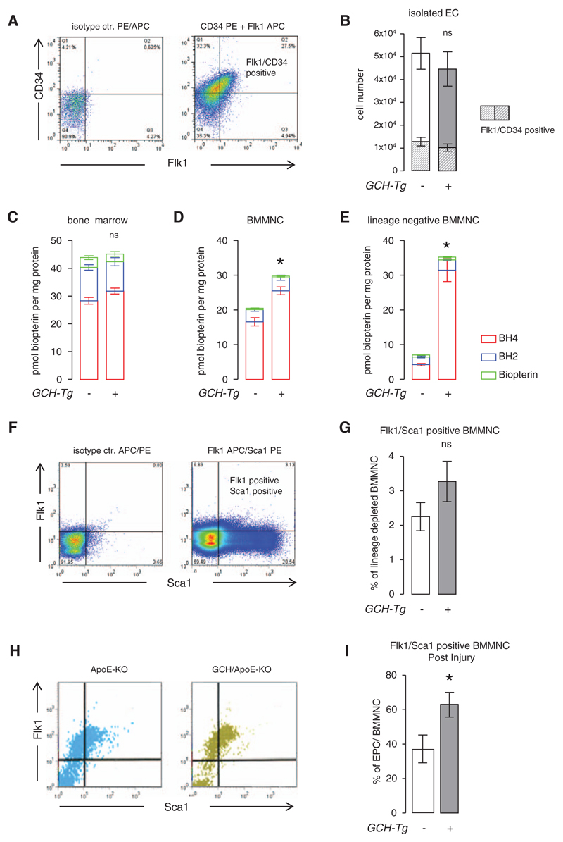
Figure 3. Endothelial progenitor cells in apolipoprotein E (apoE)-knockout (KO) and GTP cyclohydrolase (GCH)/apoE-KO mice.
A and B, The proportion of freshly isolated endothelial cells (ECs) expressing the progenitor markers Flk1 and CD34 were determined by flow cytometry. There was no significant difference in the total number of ECs isolated between the genotypes at baseline. Unpaired t test *P>0.05 GCH/apoE-KO (n=5) vs apoE-KO (n=7). Biopterin levels in bone marrow were similar (C), whereas bone marrow–derived mononuclear cell (BMMNC; D) and lineage-depleted BMMNC (E) were increased in GCH/apoE-KO mice. Unpaired t test ***P<0.001 GCH/apoE-KO (n=5) vs apoE-KO (n=7). These results indicate that in preparations of bone marrow the difference in biopterin content because of transgenic GCH overexpression becomes more apparent the higher the expected content of endothelial progenitor cell (EPC) in the sample. F–G, There was no significant difference in the percentage of Flk1/Sca1-positive BMMNC (EPC) between apoE-KO (n=11) and GCH/apoE-KO (n=9) detectable at baseline Unpaired t test *P>0.05. However, after endothelial denudation by femoral artery wire-induced vascular injury, these numbers were significantly increased in GCH/apoE-KO mice (n=11) vs apoE-KO (n=9). Unpaired t test *P<0.05. APC/PE indicates allophycocyanin/phycoerythrin.

