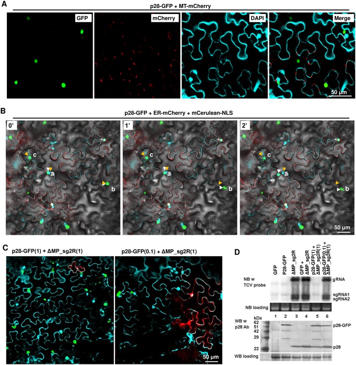
Fig 3. p28-GFP fusion protein form large, mobile intracellular inclusions that are not associated with cell nuclei or mitochondria, and repress the replication of a co-introduced TCV replicon.
Also see S3 Fig for the spatial separation of p28-GFP foci relative from ER. A p19-expressing construct was included in all infiltrations. (A) p28-GFP forms dense foci that rival nuclei (DAPI-stained) in size, and spatially separate from mitochondria and cell nuclei. MT-mCherry: a construct expressing mCherry tagged with an N-terminal mitochondrion-localizing signal. Note that DAPI stains cell nuclei as well as cell wall. (B) Mobility of p28-GFP foci. ER-mCherry: a construct expressing mCherry tagged with an N-terminal endoplasmic reticulum-localizing signal. mCerulean-NLS: a construct expressing mCerulean tagged with a C-terminal nuclear localization signal (NLS). The images additionally contained a bright field layer to help define cell boundaries. The three panels denote the same leaf area photographed at three time points: 0, 1, and 2 minutes. Three lower case letters, a, b, and c, denote three mobile p28-GFP foci, with orange and white arrowheads marking the starting and ending points, respectively. Also see a time lapse movie (S1 Video) in Supporting Information for more details. (C) p28-GFP foci do not occur in the same cells with mCherry expressed from a co-introduced replicon (ΔMP_sg2R). The two constructs were mixed at either 1:1 (left) or 0.1:1 ratios. Only merged images were presented. (D) NB and WB verifications of results in (C). Note in lane 5 that the wild-type p28 translated from the ΔMP_sg2R replicon was readily detectable despite of its inability to replicate.

