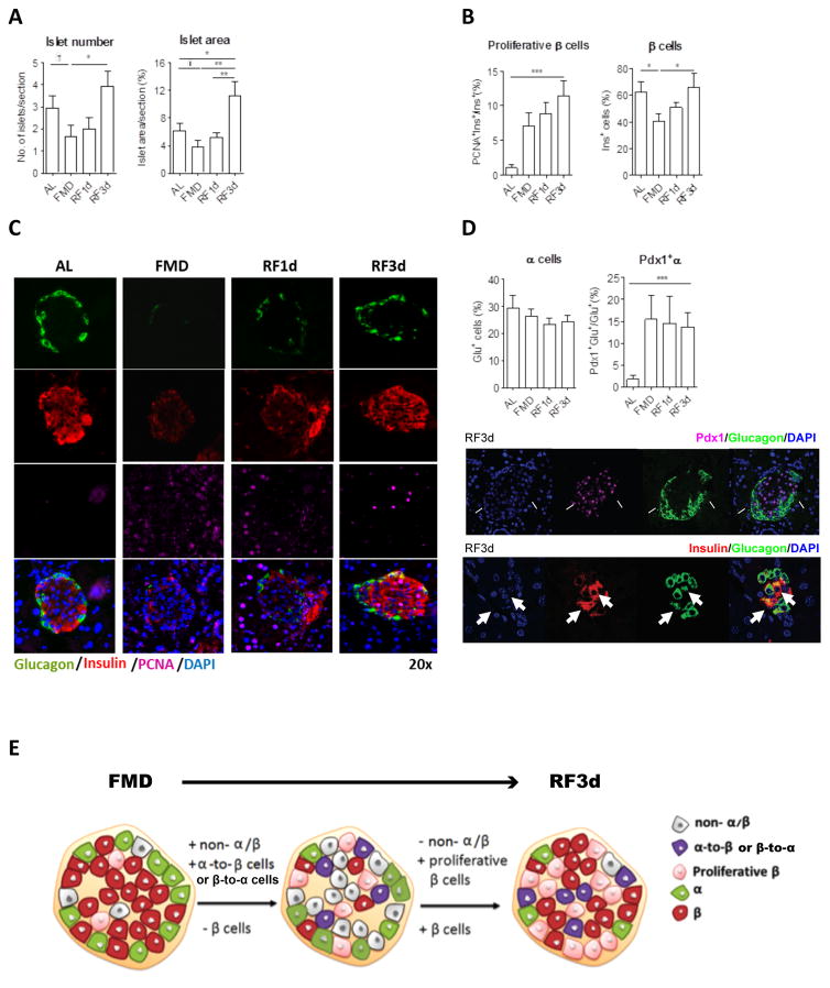
Figure 3. FMD and post-FMD refeeding promote β-cell proliferation and regeneration.
(A) Size and number of pancreatic islets per pancreatic section.
(B) Proliferative proportion of β cells and proportion of β cells per islet.
(C) Representative images of pancreatic islets with Insulin, glucagon and PCNA immuno-staining.
(D) Transitional cell population co-expressing both the markers of α and β cells: Proportion of α cells and Pdx1+α cells. Arrows in the images with split channels indicating Pdx1+Gluc+ and Insulin+Glucagon+ cells.
(E) Schematic of FMD- and post-FMD refeeding induced cellular changes in pancreatic islets.
Mice of the C57BL/6J background, at age 3–6 months, received no additional treatments other than the indicated diet. Pancreatic samples were collected from mice fed ad libitum (AL) or the fasting mimicking diet (FMD) at indicated time points: the end of the 4d FMD (FMD), 1 day after re-feeding (RF1d) and 3 days after re-feeding (RF3d); for immunohistochemical and morphometric analysis (A to E): n ≥ 6 mice per group, n ≥ 30 islets per staining per time point. mean ± s.e.m,*p<0.05, ** p<0.01, ***p<0.005, one-way ANOVA.†p<0.05, t-test.

