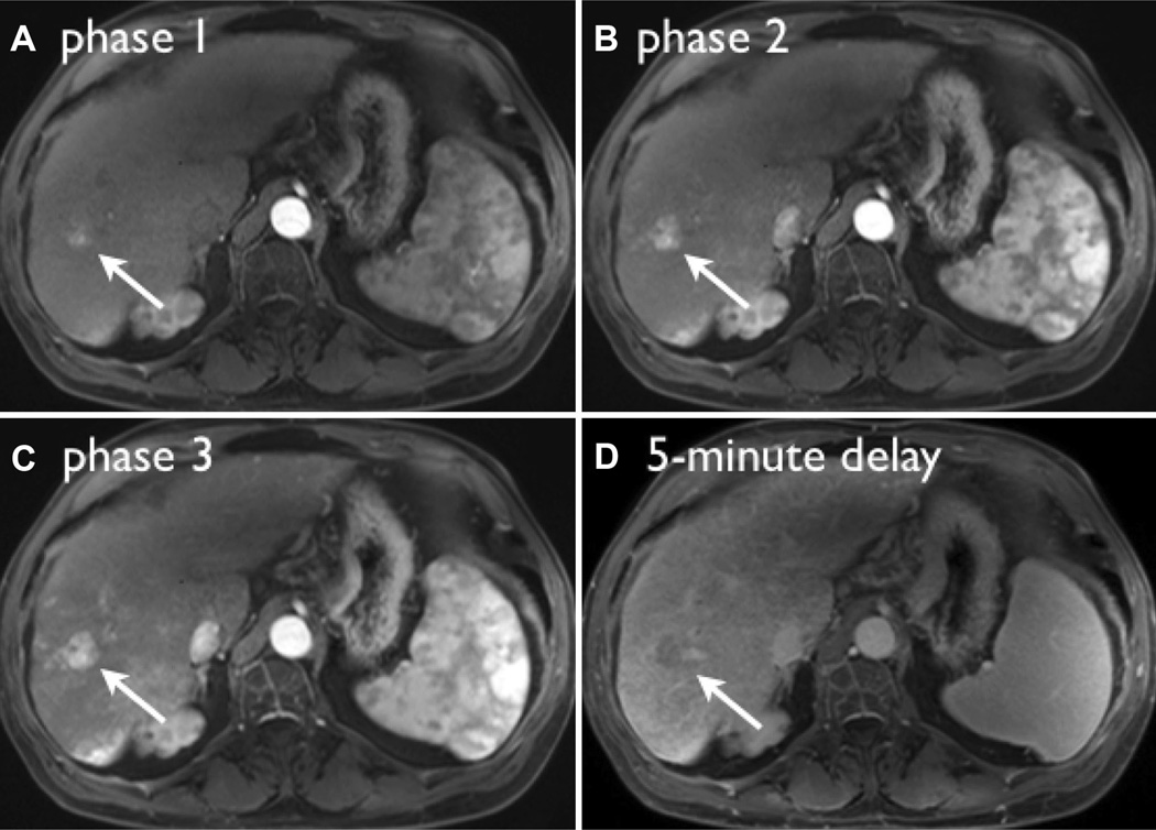
Fig. 4.
Images from representative triple-phase imaging of a LI-RADS 5 lesion, demonstrating maximum contrast-to-noise ratio (CNR) in the late arterial phase (C). The lesion demonstrates washout appearance on the 5-min delay (D). Note that in phase 1 (A), during the angiographic phase, the lesion is barely visible compared to the late arterial phase where conspicuity and lesion CNR are highest.

