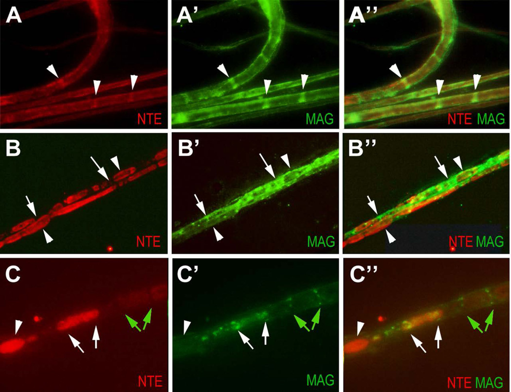
Figure 7.
NTE staining is found in ovoids along the nerve after injury. (A-A”) NTE is enriched in Schmidt-Lanterman incisures (arrowheads), co-localizing with MAG in the uninjured nerve. (B-B”). On the crushed side, strong NTE staining is detectable in ovoids along the nerve (arrowheads). NTE staining only co-localize with MAG (arrows) at the margins of ovoids. (C-C”) Magnification showing that MAG-positive incisures are adjacent to weakly stained NTE-positive ovoids (green arrows). Another ovoid that contains higher levels of NTE shows MAG-positive staining at the distal and proximal end of the ovoid (white arrows). The ovoid with the highest levels of NTE does not co-stain for MAG, nor is it delimited by MAG staining (arrowhead). The crush was performed at PND42. NTE in red, MAG in green.

