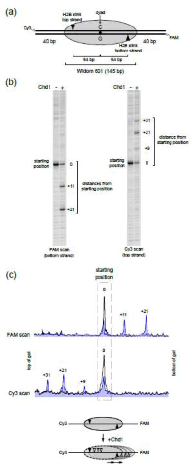
Figure 1. The Chd1 remodeler distributes Widom 601 nucleosomes asymmetrically.
(a) Diagram of a centered nucleosome on the Widom 601 positioning sequence. The orientation of the 601 sequence is defined with a dyad having a cytosine (C) on the top strand. As indicated, each H2B(S53C) cross-link occurs only on one strand.
(b) Chd1 shifts centered nucleosomes preferentially to the right side. Shown are two scans from the same remodeling reaction, which reveal the locations of H2B(S53C) cross-links for each DNA strand. For these reactions, 150 nM nucleosome was incubated plus or minus 50 nM Chd1 and 2 mM ATP for 64 min. This gel is representative of four independent experiments.
(c) Intensity scans of the gels shown in (b). The black trace represents the cross-linking distribution before remodeling, and blue trace is 64 min after addition of ATP and Chd1. Diagram below summarizes direction of octamer movement by Chd1.

