
Figure 6.
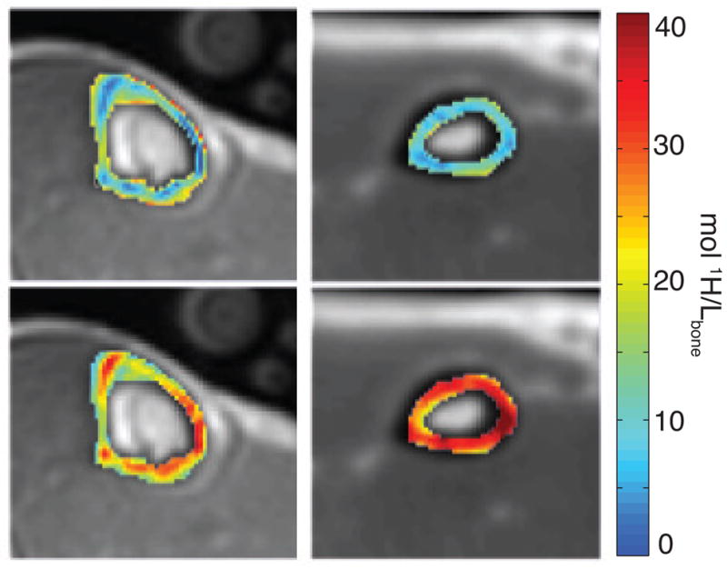
Axial MR images obtained in vivo in the lower leg (left) and wrist (right) in a healthy subject; images are conventional UTE images overlaid with pore (top row) and bound (bottom row) water maps in the tibia and the radius. Reproduced in part from (143). 45