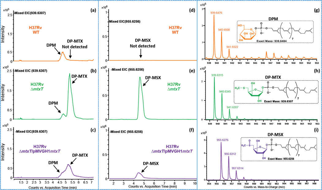
Figure 6. Negative ion LC/MS of decaprenyl-phosphoryl-sugars from total lipids extracts of M. tuberculosis H37Rv WT, the mtxT knock-out mutant and the complemented mtxT mutant strain.
Panels (a–c) are the extracted ion chromatograms (EICs) of m/z 939.6307 showing the presence of DPM (actual [M-H]− at mass m/z 939.6484; see text for details) and the presence or absence of DP-MTX in the different M. tuberculosis strains. The detection of DP-MSX at m/z 955.6256 as [M-H]− ions are shown in the EICs presented in panels d–f. The insets in the mass spectra shown in panels (g–i) show the possible structures and the exact mass of the [M-H]− ions of the decaprenyl-phosphoryl sugars detected.

