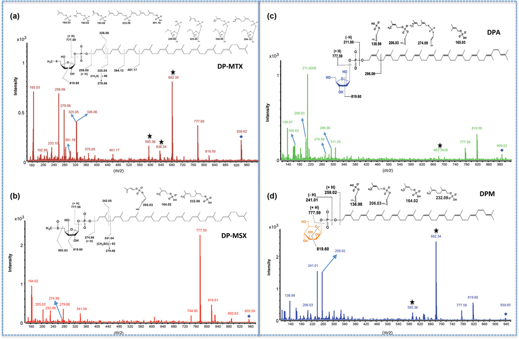
Figure 7. LC-MS/MS analysis of decaprenyl-phosphoryl sugars from total lipids extracts of the M. tuberculosis H37Rv mtxT knock-out mutant.
MS/MS spectra and proposed fragmentation patterns of DP-MTX (a), DP-MSX (b), DPA (c) and DPM (d). The fragmented precursor [M-H]− ions are identified with a diamond. The details of the cleavages are discussed in the text. Ions labeled with stars come from the fragmentation of a contaminant ion with a mass close to the m/z value of 939.6307. For DP-MTX and DPM, this ion is at m/z 939.4671.

