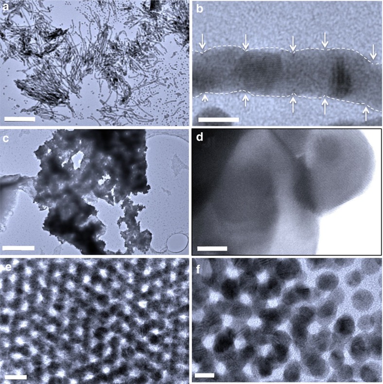
Figure 3. TEM images of consolidated gold nanostructures.
(a) Representative TEM image of the gold nanowires. (b) HRTEM image of a gold nanowire. The white dashed lines indicate the profile of individual connected gold nanocrystals. Arrows point to the interface where the gold nanocrystal coalesce. (c) Representative TEM image of gold nanosheets. (d) HRTEM image of a nanosheet with an fcc lattice fringe of (111). (e) Representative TEM image of three-dimensional gold networks. (f) HR TEM image of the three-dimensional networks showing the edge with three dimensionally connected nanocrystals. Scale bars: (a) 100 nm; (b) 5 nm; (c) 400 nm; (d) 10 nm; (e) 10 nm; (f) 5 nm.

