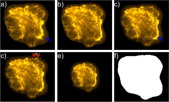
Fig. 3.

Visualization of the effect of forces employed in the energy mapping approach for a exemplary B cellcytoskeleton. The different textures in a) and c) - e) show textures with varying settings compared to texture b) simulated with settings used for evaluation: a) weaker force between bulk points resulting in stronger distortion of areas with high intensity indicated by blue arrow; c) stronger force between bulk points resulting in smaller areas with high intensity indicated by blue arrow; c) weaker force between border points resulting in serrated contour (corresponding point indicated by green arrow surrounded by serrated areas with border points indicated by red arrows); e) weaker force between corresponding points resulting in smaller texture size
