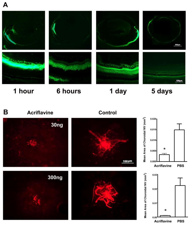
Fig 4. Acriflavine injected into the suprachoroidal is visualized throughout the entire retina and suppresses choroidal neovascularization (NV) in rats.
(A) Pigmented rats were given a suprachoroidal injection of 300 ng of acriflavine and euthanized 1 hour, 6 hours, 1 day, or 5 days after injection. Frozen ocular sections were examined by fluorescence microscopy and showed acriflavine in the anterior part of the choroid and retina on the side of the injection at 1 and 6 hours after injection. At 1 day after injection acriflavine fluorescence was greatest in the retina on the side of the injection, but could be seen throughout the posterior retina and in the retina on the side opposite the injection. At 5 days after injection, there was only faint fluorescence remaining.
(B) Pigmented rats had laser-induced rupture of Bruch’s membrane in 5 locations in each eye followed by a suprachoroidal injection of 30 or 300 ng of acriflavine in one eye and PBS in the fellow eye (n=6 for each dose). After 14 days, the rats were euthanized and choroidal flat mounts were stained with FITC-labeled Griffonia Simplicifolia lectin. A representative choroidal flat mount from an eye injected with acriflavine shows a small area of choroidal NV (left panel) compared to that from the fellow eye injected with PBS (middle panel). The mean area of choroidal NV was significantly less in eyes injected with 30 or 300 ng acriflavine compared to corresponding controls.
*p=0.0014 (30 ng); p=0.0012 (300 ng) for difference between acriflavine and corresponding PBS controls by unpaired t-test.

