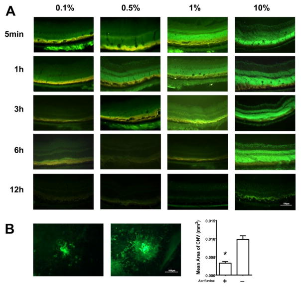
Fig 5. Topically administered acriflavine is visualized in the retina and suppresses choroidal neovascularization (NV).
(A) C57BL/6 mice had 5 μl of 0.1% (5 μg), 0.5% (25 μg) 1.0% (50 μg) or 10.0% (500 μg) acriflavine placed on the cornea of one eye of mice and 5 μl of PBS was placed in the other eye. Mice were euthanized at various times after administration and ocular frozen sections were examined by fluorescence microscopy. Fluorescence was visible in the retinal pigmented epithelium (RPE) and outer retina 5 minutes after administration of 0.1% acriflavine and was seen throughout the retina 5 minutes after administration of 0.5%, 1%, or 10%. The fluorescence became more intense and then faded with residence time in the retina greater for higher doses.
(B) Six mice had laser induced rupture of Bruch’s membrane in three locations in each eye and then had topical administration three times a day of 5 μl of 0.5% acriflavine in one eye and PBS in the other eye. After 7 days, choroidal flat mounts were stained with FITC-labelled GSA-Lectin. The mean area of choroidal NV was significantly smaller in acriflavine-treated eyes compared with PBS-treated eyes (*p=0.028 by Wilcoxon matched-pairs signed-ranks test.

