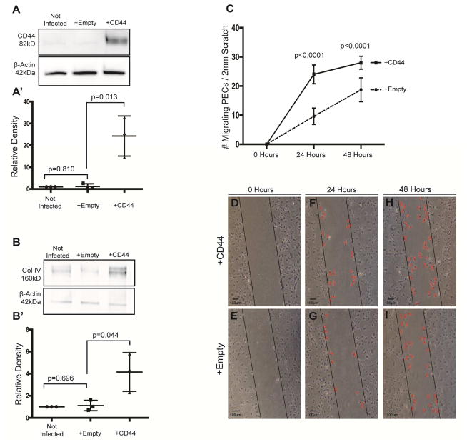
Figure 5. CD44 Infected PECs in culture have higher Collagen IV expression and increased migration.
(A) Western blot analysis for CD44. CD44 protein is not detected in non-infected immortalized mouse PECs (western blot analysis lane 1), nor in empty vector infected cells (lane 2). CD44 protein is present in cultured PECs infected with CD44 vector (third lane). The lower western blot analysis shows the protein loading control β-Actin. (A’) Densitometry confirms the significant increase in CD44 in CD44 vector infected PECs.
(B) Collagen IV western blot analysis. Low levels of collagen IV protein are detected in non-infected (lane 1) and empty vector infected (lane 2) immortalized mouse PECs. Following CD44 infection, collagen IV protein levels are increased (lane 3). The lower panel shows β-Actin used as a protein loading control. (B’) Densitometry shows that collagen IV is higher in the presence of CD44.
(C) Quantification of PEC migration. The number of cells that migrated in to the wound was quantitated in PECs infected with CD44 vector (solid line) or empty vector (dashed line). Following a scratch, PEC migration was higher in CD44 infected cells at 24h and 48h compared with empty vector infected cells. These results show that overexpressing CD44 increased the migration of cultured PECs.
(D–I) Migration assay following scratch test. Column 1 shows a representative phase contrast images of cultured mouse PECs infected with CD44 vector (upper panels) or empty vector (lower panels) following the initiation of a scratch test at time zero (D–E, column 1), 24 hours (F–G, column 2) and 48 hours (H–I, column 3). The black line is drawn to demarcate the boundary of each side of the scratch. Examples of migrating cells are indicated by a red colored X.

