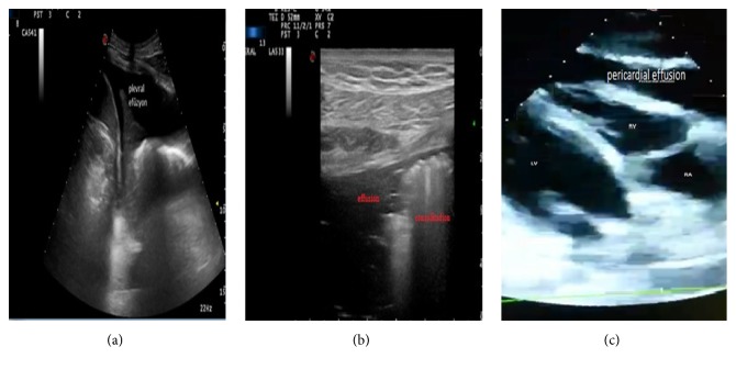
Figure 3.
Some of the ultrasonographic pathologic views obtained during the study. (a) Pleural effusion view in the left pleural area in the left upper quadrant ultrasonography. (b) Pneumonic consolidation and parapneumonic effusion view in thoracic ultrasonography. (c) View of pericardial effusion compressing the left ventricle on the parasternal scan in the cardiac ultrasound.

